Our physiotherapists are capable of treating a wide range of body issues, whether they are chronic or recent.
Discover our services and let us accompany you on your journey to healing and well-being.
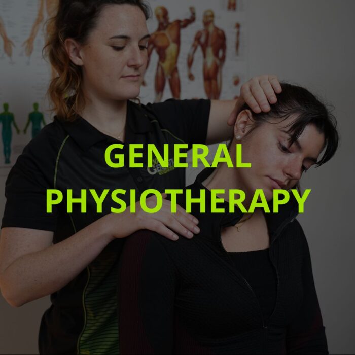
Whether it’s to recover from an injury, relieve your pain, improve your mobility, or to help you with your daily activities, we are here to help!
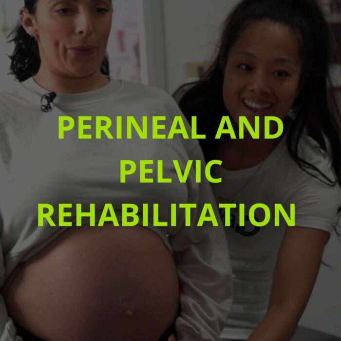
A service dedicated to the support and restoration of pelvic function, helping both women and men regain balance and comfort.

Our therapists are trained to address the unique needs of the youngest, offering care tailored to their specific requirements.

Our therapists in osteopathy, massage therapy, and acupuncture, combine their skills to optimize your overall health and prevent future injuries.
Common reasons for seeking general physiotherapy:
– Back pain due to office work: caused by long hours sitting and poor posture in front of the computer.
– Foot pain after a day spent standing: common among traders, teachers, or in the catering industry.
– Carpal tunnel syndrome in secretaries or office workers: due to excessive use of the keyboard and mouse.
– Lower back pain after lifting heavy loads at home: like lifting furniture or grocery bags.
– Tendonitis from gardening: resulting from repetitive movements when handling gardening tools.
– Knee pain after a long hike: occurring in hiking enthusiasts who may encounter rough terrain.
– Shoulder pain while painting a ceiling: caused by prolonged maintenance of arms overhead during painting.
– Ankle sprain from walking on an uneven sidewalk: a common accident in everyday life, especially on uneven ground.
– Neck pain after a long car ride: often related to a static posture during driving.
– Morning joint stiffness in the elderly: age-related.
Asking the question is answering it! As you don’t need a medical prescription, it is recommended to consult as soon as pain or discomfort is felt and your regular activities are affected.
It is clearly demonstrated that the sooner a situation is handled by a physiotherapist, the greater the chances of a quick and lasting recovery.
If you have questions, call us. We can quickly overview the situation. Then, during your first session, we will give you all the tools for a complete recovery;
– Personalized advice;
– Pain management;
– Exercise plan to support recovery.
Medial epicondylitis, also known as golfer’s elbow or Golfer’s Elbow, is a painful condition of the common tendon of the wrist flexors. This tendinopathy produces pain on the inside of the elbow, near the bony tip, as well as in the forearm and sometimes even to the wrist. It can cause a throbbing or even burning sensation inside the elbow, mainly during activities that require grasping an object.
Medial epicondylitis is primarily caused by repeated wrist and hand movements. It occurs mostly in golfers, baseball pitchers, tennis players, and manual workers. If you start a new activity of this kind or have a sudden increase in time spent doing one of them, the risk of suffering from medial epicondylitis increases. For example, this condition can occur after a golf trip or during home renovation or landscaping work.
When medial epicondylitis is quickly addressed in physiotherapy, it is possible to halt its progression and eliminate pain before it stops you from working, playing a sport, or renovating your house. Manual therapy treatments and custom exercises can accelerate the healing process, regain good joint mobility, and correct the biomechanics of arm movements.
Lateral epicondylitis, also known as tennis elbow or Tennis Elbow, is a painful condition of the common tendon of the wrist extensors. This tendinopathy produces pain on the outside of the elbow, near the bony tip, as well as in the forearm and sometimes even to the wrist. It can cause a throbbing or even burning sensation on the outside of the elbow, primarily during activities that require grasping an object, like a coffee cup, or when squeezing and twisting the hand, such as turning a doorknob or using a screwdriver.
Lateral epicondylitis is primarily caused by regular, forceful movements of the wrist and hand. It occurs especially among manual workers and tennis players. It can result from prolonged overuse, notably in construction workers, or after a sudden increase in the use of forearm muscles. For example, it can occur during home renovations or when resuming a racquet sport several times a week after a long break.
When lateral epicondylitis is quickly addressed in physiotherapy, it is possible to stop its progression and eliminate pain before it stops you from working, playing a sport, or renovating your house. Manual therapy treatments and custom exercises can accelerate the healing process, restore good joint mobility, and correct the biomechanics of arm movements.
When a limb is partially or completely immobilized following a fracture or dislocation, stiffness sets in the immobilized joints. This stiffness is due to a lack of use and is generally associated with a loss of muscle strength and control of movement. Joint stiffness and muscle weakness begin to set in from the first days of immobilization and are relatively proportional to its duration. Therefore, the longer a limb is immobilized, the more significant the consequences.
Among other things, a joint that has been immobilized for a long time can lose part of its range of motion. For example, if treatments and exercises are undertaken too late, a shoulder might not be able to lift above the head as much as the other, and a knee might not be able to extend completely. This can significantly limit sports or work activities afterward by disrupting the technique. This loss of movement will be compensated for by another region of the body, which will have to move more than necessary, placing it at risk of injury. For example, if the shoulder does not allow the arm and hand to lift an object high or hold an object above the head, the back will compensate for this lack of mobility by arching more than it should. This body adaptation can allow the desired task to be performed, but it puts the back at risk. When the gesture is made repetitively, it can cause lesions, muscle strains, and pain in the back or shoulder.
Therefore, it is important to start physiotherapy treatments and home exercises quickly to avoid the long-term consequences of immobilization on the joints. Treatments help identify the muscular and ligamentous factors causing the loss of mobility and eliminate them. Moreover, gradually adjusted exercise programs will allow you to regain the muscle strength lost during immobilization and relearn to control your movements in all their amplitudes.
A ligament sprain means that the ligament has been damaged or torn during a movement or contact. Ankle sprains and knee sprains are the most common, especially in sports involving contact and changes in direction. Several signs can indicate the presence of sprains, which are classified into three categories based on the severity of the ligament damage.
1st degree: A first-degree sprain means that the ligament has been slightly affected, but its stability is not compromised. This type of sprain is usually characterized by pain during movements that caused the sprain. There is no subcutaneous bleeding, and the presence of swelling is variable.
2nd degree: A second-degree sprain means that the ligament has been stretched and partially torn, reducing the stability of the ligament. In addition to pain during movement and at rest, the presence of subcutaneous bleeding and significant swelling is often associated with this degree of sprain, especially in the ankle. Depending on the severity of the ligament damage, a feeling of instability may be present. Physiotherapy treatments are necessary as soon as possible to accelerate healing and prevent a new sprain by restoring optimal biomechanics of the foot and ankle and improving strength and control of movements.
3rd degree: A third-degree sprain means a complete tear of the ligament. At this point, the joint loses some of its stability, and surgery is necessary to reconstruct the ligament. Third-degree sprains, if they occur during a significant impact, are often associated with other injuries, such as a meniscus tear or fracture to nearby structures, but they can also be isolated.
The vast majority of sprains can be diagnosed and treated in a clinic by a physiotherapist. However, severe sprains requiring surgery must be diagnosed by medical imaging, with an MRI or ultrasound.
It’s important to know that a ligament has little contact with the blood circulation and is therefore very limited in its repair following an injury. A stretched or partially torn ligament partially repairs itself but will never regain its previous stability. Moreover, if a significant ligament injury with the presence of bleeding, pain, and swelling is not quickly addressed, this affects the nerve receptors in your joint in the long term, thus diminishing your control over it! To avoid recurrences, it is very important to compensate for these deficits by improving the strength of your muscles surrounding the joint and training your nervous system to better manage the information coming from your joint!
The knee joint contains two menisci, the medial and lateral meniscus, which are rings of fibrocartilage that improve the stability of the knee and reduce pressure on the bones. Several types of injuries can occur to the menisci. Magnetic resonance imaging can show a small cut in the ring, up to a complete tear of a portion of the meniscus and even the presence of pieces floating in the joint. A meniscus can be damaged traumatically following a contact or a sprain, but it can also suffer progressive lesions over the years. The menisci receive little blood circulation, and their recovery is very limited. Their healing following an injury varies depending on the injured section, which is why the most common meniscal surgeries involve removing a part of the meniscus.
However, although the section of the meniscus is removed following this type of surgery, the disappearance of pain is not guaranteed. Moreover, the stability and biomechanics of the knee are affected. This increases pressure and friction on the bone surfaces in the knee and raises the risk of early onset osteoarthritis. Knee meniscal surgery should be considered mainly if the meniscus blocks or limits movement after moderate physiotherapy treatment.
Moreover, although the precise diagnosis is made by medical imaging, it is important to know that several clinical signs allow the physiotherapist to identify whether a meniscus is affected or not. Therefore, an MRI is not necessary to evaluate and treat your knee if you think you have suffered a meniscal injury. A torn meniscus would cause a sensation of blockage in the knee during certain movements like going downstairs, sitting on a chair, or doing a squat.
Knee pain during meniscal injury is mainly produced by the bones pressing on the injured section of the meniscus or by stretching it. However, the menisci are closely linked to the ligamentous and muscular structures surrounding the knee and can sometimes be trapped by poor joint alignment or improper movement. It is often possible to correct the alignment and movement to reduce the pressure or trapping of the injured meniscus section. Therefore, if you think your meniscus is affected, know that it is not necessary to immediately consult in medicine or undergo medical imaging tests to start treatments. With quick physiotherapy management, it is possible to eliminate knee pain and limit the progression of the lesion to avoid surgery.
Groin pain can be caused by several types of injuries. It can occur due to muscular issues such as strains and sprains or due to a lack of mobility in the hip joint, among other reasons. A sensation of pinching in the groin can occur when the hip is bent or when the leg is crossed over the other in a seated position. This type of pain often occurs in hockey players due to the type of muscular effort required by skating, which causes a lot of tension in the small stabilizing muscles of the hip. These muscles, when tense, limit the mobility of the hip and cause a change in the movement of the joint.
The hip joint is spherical, allowing movements in all directions. Muscular tensions can misalign the joint and cause excessive friction of the femoral head, which is round, on the anterior part of the acetabulum, the cavity in the pelvic bone that receives the round head of the femur. This round cavity in the bone also has a cartilaginous rim called the labrum, which helps stabilize the head of the femur in the cavity. This can sometimes get trapped between the bones and cause pain. Similar to the menisci in the knee, it is possible to restore flexibility and mobility to the hip to regain optimal biomechanics of movement that does not cause pain.
Other types of injuries can also cause groin pain, including back problems. If groin pain is combined with other pains, either in the knee, back, or genital area, it is important to consult a physiotherapist or doctor to eliminate the possibility of serious conditions.
A muscle strain typically occurs during an intense effort coupled with stretching of the affected muscle. It is actually a stretch or tear of the muscle with varying severity. It causes a snapping sensation and sharp pain, forcing an immediate stop to the activity due to the pain. Muscle strains are classified into three categories.
The first grade involves stretching of the muscle with damage to muscle cells, but without tearing. Pain will be present during movement when contracting and stretching the muscle. A loss of strength may be present.
The second grade involves a partial muscle tear, usually visible on a muscle ultrasound. A decrease in strength and sharp pain when stretching the muscle will be evident. Depending on the severity and type of tear, surgery may be necessary shortly to reattach the sections of the muscle and allow healing.
Finally, the third grade is a complete tear of the muscle. At this point, significant or total loss of strength is noted along with significant subcutaneous bleeding. Surgery is then necessary immediately to reattach the sections of the muscle and allow healing. This is a medical emergency, and surgery should be performed within hours of the tear for optimal results.
The signs of strain mentioned above, such as pain or reduction in movement and strength, are often proportional to the severity of the strain. In all cases, relative rest should allow protection of the muscle to avoid increasing muscle damage. This rest is not complete and should include circulatory exercises to maintain good blood circulation in the affected area and promote healing of the injury. It is very important to treat a muscle strain to avoid weakening of the muscle and compensation by other muscles or the opposite side (especially for the legs). This endangers other muscles and increases the risk of recurrence in the same muscle upon returning to sport.
Orthopedic surgeons trust physiotherapists as health professionals and movement specialists to help you regain your mobility, strength, and function following surgery. Whether it’s after a fracture, the implantation of a prosthesis, or any other orthopedic surgical intervention, prompt physiotherapy management can speed up the recovery of joint mobility and adjust exercises for optimal progression.
Following surgery, physiotherapy services are offered by the public health system. However, these services are limited in availability and are mainly focused on regaining independence at home and safety in everyday movements. Regardless of your age and the type of surgery undergone, it is important for your health to continue physical activity and sports after surgery. However, the risk of new injuries is increased after surgery due to decreased muscle strength and limitations in range and control of movements. That is why special attention must be paid to restoring normal muscle functions for a return to sport.
Our expertise in sports physiotherapy allows us to help you progress more quickly after surgery and guide you safely in resuming your work or sports activities.
A herniated disc is a condition affecting an intervertebral disc that acts as a cushion between each vertebra. This condition is more commonly present in the neck and lower back. It causes painful symptoms locally and sometimes radiating to the arms or legs. Among other things, the presence of a feeling of stiffness in the lower back preventing straightening up may be a sign of a herniated disc. Being in a lying position generally relieves this pain compared to standing or sitting, as it reduces the pressure exerted by the upper body on the intervertebral disc.
A herniated disc can also cause various neurological symptoms such as tingling or numbness in the limbs and a decrease in muscle strength or skin sensitivity. These symptoms may indicate a medical emergency if they correspond to compression of the spinal cord and should be evaluated by a physiotherapist or a doctor. If you notice the presence of such symptoms bilaterally or simultaneously with urinary or fecal disturbances, a decrease in sensitivity in the genital area, or significant weakness in an entire limb, this is a medical emergency. This situation requires a visit to the emergency room.
The symptoms of a herniated disc are often similar to those of a lumbar strain or sciatica. A combination of these various conditions may also occur. Clinical assessment in physiotherapy allows evaluating the need for a medical consultation and magnetic resonance imaging. Various treatments tailored to your condition will help control and relieve pain while providing you with the best tools to recover good mobility of the back and optimal posture.
Lower back pain, also known as lumbalgia, can be caused by a multitude of conditions. It may result from a specific event, such as lifting a heavy box, or develop gradually over several months or even years. The term “lumbalgia” is very broad and encompasses a variety of painful conditions that can be caused by both tense muscles and a lack of mobility in the spinal joints, often a combination of several factors. Physiotherapists specialize in assessing and treating all these different factors that can cause your back pain. Additionally, posture, whether at work, during leisure activities, or at home, as well as sleeping positions, can significantly influence back pain. Finding an appropriate sleeping posture can improve your night’s sleep and greatly help reduce your back pain.
Sciatica refers to a painful condition related to the sciatic nerve, which originates in the lumbar spine and has branches that extend down to the foot. Thus, certain back conditions may be accompanied by pain radiating down the leg, with sensations of numbness, burning, or heaviness. Posture, both at work and at home, muscle tension, as well as a lack of mobility and control of movement in the back and hips, are primarily responsible for painful back conditions. Back pain affects manual laborers, office workers, and athletes alike. Our expertise in physiotherapy allows us to target all the causes of your back pain and treat them, relieving your pain and preventing its recurrence.
Back pain and sciatica often occur for the first time long before they become bothersome at work or in our leisure activities. The pain is often occasional, bearable, or endurable for a while until the day when it is no longer so. Thus, even if some back pain disappears with rest, the mobility and posture problems causing this pain do not vanish over time. These problems accumulate and worsen, eventually leading to changes in movement habits and physiological changes in tissues and nerve connections. This, in the long run, causes the blockages, pinches, and pain felt at work, after a workout, or during prolonged standing or sitting, such as during a social gathering or a car trip, for example.
Therefore, it is important to address back pain promptly and learn to resolve it rather than learning to live with it. It is not optimal to simply treat the pain without addressing the underlying causes or making changes in your lifestyle habits. Back pain is not inevitable or unchangeable. Our vision is to teach you how to take care of your back health yourself, every day.
Tendonitis and tendinopathy affect tendons, which are the fibrous part at the end of a muscle that attaches to the bone. All tendons can be affected, so the term “tendonitis” is often associated with the name of the tendon or muscle in question. However, there are significant differences between tendonitis and tendinopathy, which will be outlined here.
Tendonitis is an inflammatory condition affecting a tendon as a result of its overuse over a short period. For example, Achilles tendonitis can occur in a novice runner who starts with too much training volume and intensity. It is often accompanied by limitations in joint mobility, muscle imbalances, or suboptimal technique in the activity. Tendonitis is primarily felt as pain and a sensation of burning or throbbing, present during movement when the muscle contracts and when it stretches. This pain is often more intense in the morning upon waking and after provocative activity. Ice and anti-inflammatories help in the short term to alleviate pain, but scientific evidence is mixed on their long-term impact on tendon healing. Relative rest in activities that increase pain, along with a gradual return to these activities following specific mobility and strengthening exercises for the affected tendon, allows for a quicker return to sports or work with less risk of retaining pain than complete rest with anti-inflammatories.
Tendinopathy differs from tendonitis in that it is not inflammatory. It is a more long-term and severe wear and tear of the tendon that can occur if tendonitis becomes chronic or simply due to poor or overuse of the tendon over time. In this situation, the tendon transforms, and its fibers lose their optimal alignment. For example, rotator cuff tendinopathy in the shoulder, often referred to as shoulder tendonitis, occurs due to overuse of these tendons over the years and is mainly associated with shoulder impingement syndrome and posture. In this type of condition, the tendons are overused and are trapped between the scapula and the humerus, increasing damage to the tendon. Tendon degeneration can lead to cracks in the tendon and eventually complete rupture.
The treatment of tendinopathy mainly involves pain management and correction of movements causing overuse of the tendon. Therefore, it is important to work on the mobility and biomechanics of the involved joints, as well as to improve muscle strength, movement control, and tendon resistance with specific exercises. For example, for the shoulder, correcting posture and movements involving raising the arms helps reduce the load on the affected tendons. When combined with specific exercises to better control the scapula and strengthen the rotator cuff, pain decreases significantly without the need for medication. Moreover, all activities requiring holding an object in the air or lifting a load become easier.
Une bursite est une inflammation d’une bourse, qui sert de coussin entre une surface osseuse et des tendons. Ces bourses se situent généralement au niveau d’une articulation comme à l’épaule ou au genou par exemple. La plus courante est la bursite d’épaule, qui ressemble beaucoup à une capsulite, mais qui évolue beaucoup plus rapidement et dont les symptômes sont soulagés en quelques jours par des anti-inflammatoires et l’application de glace.
Une bursite peut survenir à la suite d’un trauma précis ou à la suite d’une surutilisation et des microtraumatismes répétés. Par exemple, la bursite de l’étudiant est une inflammation de la pointe du coude. Elle est nommée ainsi puisqu’elle survient principalement chez les étudiants qui passent de longues heures à lire durant la période d’examens avec la tête appuyée sur leur main et le coude sur la table!
Les bursites peuvent évoluer en bursopathies lorsqu’elles ne sont pas prises en charge, ni traitées. À ce moment, des douleurs persistantes et une limitation dans certains mouvements peuvent survenir.
Frozen shoulder is a shoulder condition that evolves slowly and quite characteristically in three phases, lasting an average of 30 months in total. The first is the painful phase, followed by the adhesive phase, and finally the regressive phase. Although its progression is slow, frozen shoulder leaves few functional sequelae, and it is extremely rare to experience a recurrence in the same shoulder. This condition more often affects women than men and typically occurs between the ages of 50 and 70, mainly in the non-dominant shoulder. While it has a favorable natural evolution, frozen shoulder remains highly inconvenient, and several solutions can reduce its impact on pain and function.
Pain appears first, mainly at night but also during the day, both at rest and during shoulder movements, without clear factors increasing or triggering this pain. This phase can last from 2 to 9 months and generally does not present limitations in movement. At this stage, the main goal is to achieve optimal mobility, strength, and stabilization in the shoulder and to address any other issues that may cause pain and be misdiagnosed as frozen shoulder. Early physiotherapy intervention helps reduce stiffness and shorten recovery time in the following phases. Taking analgesics helps control pain and improves tolerance to exercises.
Subsequently, stiffness gradually sets in along with pain during the adhesive phase of frozen shoulder, thus limiting all shoulder movements. It is usually at this point that the diagnosis is made, as this is when frozen shoulder most significantly restricts activities, prompting individuals to seek healthcare professionals. It becomes almost impossible to lie on the affected shoulder and perform large amplitude movements with the shoulder. This phase significantly limits activities and can last from 3 to 9 months if not addressed. Corticosteroid injections in the shoulder, combined with physiotherapy treatments consisting of exercises, specific stretches, and precise joint mobilizations, help improve movement ranges and the ability to use the arm more quickly. Analgesics are still used for pain management.
Finally, the regressive phase begins when pain gradually diminishes and joint stiffness improves. This phase can last a total of 12 to 42 months, but the increase in mobility occurs gradually during this period. Continuing exercises and treatments accelerates progress. There must also be a resumption of strength and muscle control in ranges that have not been reached for several months.
The cessation and resumption of daily and sports activities are based on the progression of shoulder mobility, pain, and strength. Although this condition is highly inconvenient, especially in the adhesive phase, complete rest is strictly discouraged as it increases stiffness. It is important to maintain activities according to tolerance and to perform prescribed physiotherapy exercises to avoid other complications, both physical and psychological, throughout the entire duration of frozen shoulder.
La cervicalgie est une condition douloureuse qui est causée par la région du cou, mais qui peut être ressentie de différente façon, autant par des maux de tête que des douleurs locales au cou et dans le haut du dos. Une combinaison de douleurs peut aussi survenir à certains moments de la journée ou durant certaines activités. Ces douleurs sont principalement causées par des tensions musculaires, un manque de mobilité entre les vertèbres ou un coincement des nerfs à leur sortie de la colonne vertébrale. La posture a un rôle extrêmement important dans les douleurs au cou. En effet, une mauvaise posture du dos et du cou augmente beaucoup la tension musculaire dans le cou et diminue la quantité de mouvements disponibles.
Une cervicalgie peut provoquer des douleurs dites mécaniques, apparaissant au mouvement et disparaissant au repos. Typiquement, elles seront ressenties comme un pincement ou un blocage lors des mouvements de la tête, en regardant les angles morts en voiture ou le moineau lors d’une partie de badminton par exemple. Lorsque répétés plusieurs fois, ces mouvements peuvent provoquer une irritation de la section du cou qui manque de mobilité et provoquer une augmentation des symptômes ou l’apparition de nouvelles douleurs. Ainsi, un pincement tolérable dans le cou en début de partie de badminton peut devenir très douloureux vers la fin du match et même provoquer des maux de tête ou des douleurs descendantes au niveau de l’épaule et du bras.
La cervicalgie peut aussi provoquer des douleurs dites neurologiques, qui sont plutôt ressenties comme des sensations de brûlures, de chaud/froid ou de décharges électriques. Ce type de douleur a tendance à augmenter au courant de la journée ou de l’activité et avec la fatigue et le stress. Il est important de différencier une brachialgie avec douleurs neurologiques d’une hernie discale cervicale. Si vous ressentez des douleurs, une perte de force ou une diminution de la sensibilité de la peau en même temps dans les deux bras ou dans les jambes, il est important de consulter un physiothérapeute ou un médecin le plus rapidement possible. Il est aussi important de consulter rapidement si ces douleurs sont accompagnées de vertiges, de troubles de vision ou d’audition, de pertes de conscience ou de migraines.
La cervico-brachialgie implique des douleurs combinées au cou et dans le bras ou uniquement au bras, mais provenant du cou. Tel que mentionné plus haut, une douleur peut provenir du cou et descendre dans le bras lorsqu’augmentée, mais peut aussi être ressentie uniquement dans le bras, notamment lors de compressions nerveuses. En effet, les nerfs du bras débutent au niveau du cou à leur sortie de la colonne vertébrale et se rendent jusqu’à la main. C’est pourquoi des douleurs au cou peuvent être accompagnées de douleurs n’importe où dans le bras et l’épaule en fonction des nerfs atteints.
Les traitements de physiothérapie permettent notamment de réduire la douleur et les tensions musculaires, de retrouver la mobilité au niveau de votre cou et de vous aider à corriger votre posture. Nous voulons vous donner les outils nécessaires pour que vous puissiez mieux comprendre ce qui provoque vos douleurs, comment les éliminer et comment éviter qu’elles reviennent.
The carpal tunnel syndrome occurs when there is compression of the median nerve as it passes through the carpal tunnel at the wrist. This syndrome primarily causes numbness in the hand and sometimes pain in the thumb and fingers, which can manifest as intense burning sensations or a feeling of cold in the hand. It is often more troublesome at night and during manual activities, potentially leading to a loss of strength and difficulties in gripping certain objects.
Carpal tunnel syndrome can be caused by a restricted space in the carpal tunnel or physiological changes resulting from many years of manual activities. It may also be associated with other nerve compressions along its course between the spine and the wrist, thereby increasing its sensitivity to compression within the tunnel. Consequently, it is useful to address both the elbow, shoulder, back, and forearm to prevent double-crush syndrome. Physiotherapy treatments are often effective in avoiding more invasive interventions, such as surgery.
The use of an immobilization brace, especially at night, is beneficial in the short term to alleviate symptoms. However, if symptoms persist for more than 6 months and conservative treatments are ineffective, nerve decompression surgery may be considered.
Plantar fasciitis is characterized by pain under the foot, especially during the first steps in the morning and during walking or running. It involves an inflammation of the plantar fascia, which is a broad fibrous band under the foot that, along with the foot muscles, supports the arch of the foot. The arch of the foot is the curvature between the heel and the toes, necessary for absorbing shocks during walking, running, and jumping. Thus, during walking, the arch flattens, stretching the plantar fascia and causing pain. This condition develops gradually and occurs more frequently in runners and individuals with flat feet.
Plantar fasciitis can occur due to changes in footwear, training volume, or running technique. For instance, if the previous running shoe provided arch support, and the new one is flatter, it alters the foot’s biomechanics and the tension in the fascia during running. It can also occur in individuals who spend a significant part of the day standing or walking, even without regular training.
Although orthotics are often prescribed to address the situation, it is possible to treat plantar fasciitis by correcting the biomechanics of the foot and leg. Indeed, with relative rest from painful activities, foot and leg relaxation and strengthening exercises, and the use of orthotics as a crutch to support the arch during walking. They help reduce tension on the fascia but do not correct its degeneration or the mechanics involved.
This syndrome encompasses a wide range of issues related to the movement of the patella (kneecap) on the femur (thigh bone). Primarily, there is pain in front of or slightly outside the patella. The pain is mainly experienced during certain activities such as descending stairs, running, swimming, and hiking. It may occur early, from the first steps, or only after the 10th kilometer, but generally at the same point each time. The same pain can also be triggered by maintaining a specific sitting position for an extended period, such as during a school class or a car journey, for example. The sitting position causing pain varies among individuals as the pressure points between the patella and femur change depending on the knee position. Thus, two people with femoropatellar syndrome may have different tolerances, with one unable to endure long-distance driving, and the other unable to sit upright in a chair for an extended period without stretching their legs.
Generally, femoropatellar syndrome is associated with muscular imbalance in the thigh and buttocks. This imbalance increases pressure and friction between the patella and femur, leading to irritation and knee pain. These imbalances may also be accompanied by joint stiffness or postural factors altering the alignment of the knee or hip. Additionally, there is an increased risk of developing femoropatellar syndrome following a knee injury or the use of suboptimal techniques in sports activities, such as running, for example.
Rest, the use of anti-inflammatory medications, and ice can help reduce irritation and control pain over a short period but are not a medium to long-term solution. It is important to identify the factors causing femoropatellar syndrome and address these issues to prevent more serious damage to the bone surface. This involves rehabilitation over several weeks with specific strengthening and movement control exercises, combined with relative rest from painful activities. The return to sports and painful activities should be gradual to allow the body to adapt to new movement techniques.
Mainly present in runners, iliotibial band syndrome causes debilitating pain on the outside of the knee and sometimes on the side of the thigh during physical activity. The iliotibial band is a fibrous band on the side of the thigh that starts at the hip and extends down to the knee. The band syndrome occurs due to friction of the band during movement, on the outer point of the knee bone. When done repeatedly, this friction irritates the band and can even cause swelling around the affected area. This irritation can be triggered by a sudden change in the volume and intensity of training or by a change in surface or terrain in cyclic sports such as running and cross-country skiing.
While some hereditary anatomical factors may predispose someone to iliotibial band syndrome, the main causes leading to the onset or worsening of symptoms are related to muscular imbalances, joint stiffness, sports technique, and training management. For example, weakness in the gluteal muscles, combined with an imbalance between the outer and inner parts of the quadriceps and a lack of control of the leg when it hits the ground during running, is often observed in runners with iliotibial band syndrome. It is crucial to identify the biomechanical and muscular factors that predispose the athlete to iliotibial band syndrome and to pinpoint recent changes in training.
Treating the factors causing irritation of the band on the bone, combined with relative sports rest and a gradual return to activity, helps eliminate symptoms. This approach also allows for a quicker return to training with less loss of performance than complete rest.
We believe in a proactive approach. Consulting a physiotherapist early can not only reduce pain, but also significantly increase your chances of a quick and lasting recovery. During your first session with us, expect to:
– Receive personalized advice tailored to your situation.
– Learn strategies to effectively manage pain.
– Get an exercise plan designed specifically to support and accelerate your recovery.
Do you have questions or concerns?
